Progress has been made in joining bone fragments that once formed the inner linings of many of the pipes. To date, 11 bulbs (mouth pieces) and 11 reed holders have been identified and assembled. Both of these sections exhibit a surprising variety of sizes and surface decorations, supporting the notion that the pipes also varied in size and shape. It is for now certain that at least 11 pipes once existed.
Below are the 11 bulbs, largely reconstructed from fragments. There are four additional short cylindrical bone sockets, which may once have connected bulbs into the bronze tubing of an instrument. Whether these sockets belong to any of the 11 bulbs or if they point to an even larger number of bulbs, and therefore pipes, remains an open question at the moment as no direct join has been identified.

The bulb below is quite well preserved and almost intact; it still carries a once shiny and decorative silver sleeve, which is now corroded and brownish in color.

Below are the 11 identified and partially reconstructed bone bells. A few are still covered with the thin bronze that enveloped their exterior. A large number of bronze pieces remain to be joined.

On this reed holder, the inner bone lining is still encased by an extremely thin bronze sheet (less than 0.5 mm thick), with an inwardly turned edge that is perfectly fitted around the bone.

X-ray Fluorescence
Surface analysis through X-ray fluorescence of various elements of the instruments, including straight tubes, knobs, and metal decorations on the bone bulbs, has provided initial insights into the metals used in their manufacture. It appears that large sections of the round tubes were made from a copper tin bronze. Lead was detected in areas which were once joined by soldering, and silver was found on various bone bulbs.
X-radiography
To date, all metal elements have been examined by X-radiography. Radiographs allow visualization of the highly corroded metal parts, as well is viewing of internal features, not readily visible to the naked eye. X-ray images also aid in understanding how pieces were originally made, identifying for example joins and solder seams. The radiographs also give insight into the highly sophisticated fine mechanics of the auloi: uniform sections of extremely thin and straight bronze tubing fitted into each other with less than a tenth of a millimeter gap.

A radiograph of the bone bulb (hypholmion) pictured above reveals a perfectly straight and uniform borehole in its interior. The white section corresponds to a decorative silver sleeve, which is more radio-opaque than the bone.
The radiograph below shows a triple tube with bone lining and two bronze sleeves with bronze knobs attached. The knobs allow the outer sleeve to be rotated around the inner, enabling the player to switch between different musical modes as well as to move between different pitch regions.

Below, juxtaposition of a complex corroded tube with its radiograph illustrates how much information is revealed by this type of examination.

The radiograph of this round tube section shows a rectangular shadow around a round hole. Further examination by X-ray fluorescence confirms that this shadow corresponds to the presence of the residue of a lead-rich solder.
The inclined bronze tube below was soldered onto a pipe during manufacture (see above). Over time, the solder corroded and the once connected items became detached.

Scanning Electron Microscopy (SEM)/Energy-dispersive X-ray Spectrometry
A number of segments were small enough to be directly examined in the chamber of the SEM and did not require sampling.
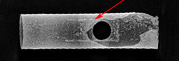
This back-scattered electron image shows tin-rich islands on the surface of the inclined bronze tube pictured above. The tin-rich surface may be a phenomenon caused by corrosion, or may possibly suggest original and intentional tinning of the surface. Further examination is required to determine the exact nature of this surface.

In a number of rotating sleeves, remnants of textile fibers were found. The back-scattered electron image below clearly shows a twisted yarn.
